
Acquisition of EEG signals using biomedical systems
Introduction
The electroencephalogram (EEG) is the recording of the brain’s electrical activity, obtained by measuring the biopotentials generated by neuronal activity at the surface of the scalp or directly within brain tissue. Recorded using surface electrodes placed on the scalp, or in some cases using needle electrodes inserted into brain tissue.Generated by the electrical activity of neurons in the brain, resulting mainly from postsynaptic potentials of large groups of cortical cells. The signal amplitude typically ranges from 10 to 100 μV (peak). Its frequency content extends from DC up to approximately 100 Hz.
The Nervous System
The task of controlling the various functions of the body and coordinating them into an integrated living organism is not simple. Consequently, the nervous system, which is responsible for this task, is the most complex of all systems in the body. It is also one of the most interesting. Composed of the brain, numerous sensing devices, and a high-speed communication network that links all parts of the body, the nervous system not only influences all the other systems but is also responsible for the behavior of the organism. In this broad sense, behavior includes the ability to learn, remember, acquire a personality, and interact with its society and the environment. It is through the nervous system that the organism achieves autonomy and acquires the various traits that characterize it as an individual.Anatomy of the Nervous System
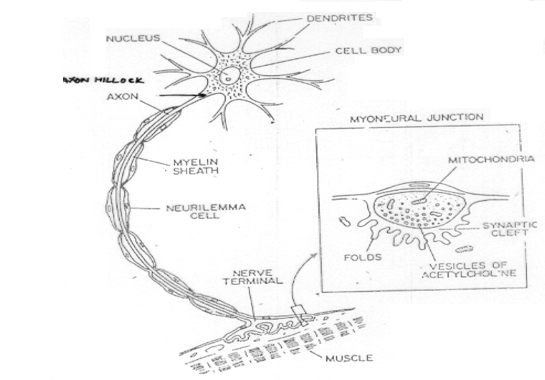
The basic unit of the nervous system is the neuron. A neuron is a single cell with a cell body, sometimes called the soma, one or more “input” fibers called dendrites, and a long transmitting fiber called the axon. The portion of the axon immediately adjacent to the cell body is called the axon hillock. Both axons and dendrites are called nerve fibers, and a bundle of individual nerve fibers is called a nerve. Nerves that carry sensory information from the various parts of the body to the brain are called afferent nerves, whereas those that carry signals from the brain to operate various muscles are called efferent nerves.
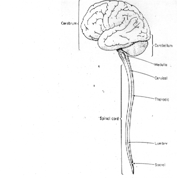
The brain is an enlarged collection of cell bodies and fibers located inside the skull, where it is well protected from light as well as from physical, chemical, or temperature shock. At its lower end, the brain is connected with the spinal cord, which also consists of many cell bodies and fiber bundles. Together the brain and spinal cord comprise one of the main divisions of the nervous system, the central nervous system (CNS). In addition to a large number of neurons of many varieties, the central nervous system also contains a number of large fatty cell bodies called glial cells. About half the brain is composed of glial cells.
Cell bodies and small fibers in the fresh brain are grey in color and are called grey matter, whereas the myelin coating of larger fibers has a white appearance, so that a collection of these fibers is referred to as white matter. Collections of neuronal cell bodies within the central nervous system are called nuclei, while similar collections outside the central nervous system are called ganglia.
Nerve fibers outside the central nervous system are called peripheral nerves. This name applies even to fibers from neurons whose cell bodies are contained within the central nervous system. Throughout most of their length, many peripheral nerves are mixed, in that they contain both afferent and efferent fibers. Afferent peripheral nerves that bring sensory information into the central nervous system are called sensory nerves, whereas efferent nerves that control the motor functions of muscles are called motor nerves. Peripheral nerves leave the spinal cord at different levels, and the nerves that innervate a given level of body structures come from a given level of the spinal cord.
The peripheral nervous system actually consists of several subsystems. The system of afferent nerves that carry sensory information from the sensors on the skin to the brain is called the somatic sensory nervous system. Visual pathways carry sensory information from the eyes to the brain, whereas the auditory nervous system carries information from the auditory sensors in the ears to the brain. Fig.2 shows a side view of the brain and spinal cord, and Fig.3 is a cutaway showing some of the major structures.
Human Brain
The human brain is an amazing three-pound organ that controls all functions of the body, interprets information from the outside world, and embodies the essence of the mind and soul. Intelligence, creativity, emotion, and memory are a few of the many things governed by the brain. Protected within the skull, the brain is composed of the cerebrum, cerebellum, and brainstem. The brain receives information through our five senses: sight, smell, touch, taste, and hearing - often many at one time. It assembles the messages in a way that has meaning for us, and can store that information in our memory. The brain controls our thoughts, memory and speech, movement of the arms and legs, and the function of many organs within our body.
Brain Region
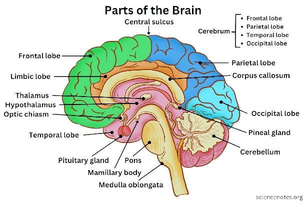
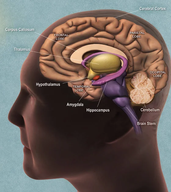
The cerebrum is the largest brain structure and part of the forebrain (or prosencephalon) (Fig.4). Its prominent outer portion, the cerebral cortex, not only processes sensory and motor information but enables consciousness, our ability to consider ourselves and the outside world. It is what most people think of when they hear the term “grey matter”. The cortex tissue consists mainly of neuron cell bodies, and its folds and fissures (known as gyri and sulci) give the cerebrum its trademark rumpled surface. The cerebral cortex has a left and a right hemisphere. Each hemisphere can be divided into four lobes: the frontal lobe, temporal lobe, occipital lobe, and parietal lobe. The lobes are functional segments. They specialize in various areas of thought and memory, of planning and decision making, and of speech and sense perception.
Lobes of the Brain
Brainstem acts as a relay center connecting the cerebrum and cerebellum to the spinal cord (Fig.4). It performs many automatic functions such as breathing, heart rate, body temperature, wake and sleep cycles, digestion, sneezing, coughing, vomiting, and swallowing. The brain stem connects the spinal cord to the higher-thinking centers of the brain. It consists of three structures: the medulla oblongata, the pons, and the midbrain. The medulla oblongata is continuous with the spinal cord and connects to the pons above. Both the medulla and the pons are considered part of the hindbrain. The midbrain, or mesencephalon, connects the pons to the diencephalon and forebrain. Besides relaying sensory and motor signals, the structures of the brain stem direct involuntary functions. The pons help control breathing rhythms. The medulla handles respiration, digestion, and circulation, and reflexes such as swallowing, coughing, and sneezing. The midbrain contributes to motor control, vision, and hearing, as well as vision- and hearing-related reflexes.
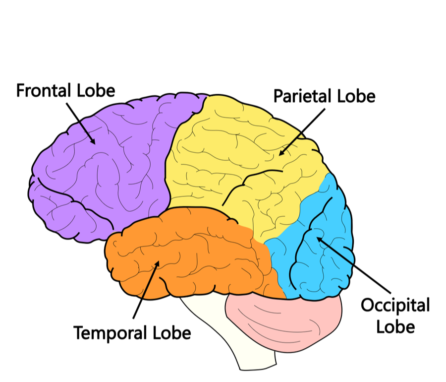
- Frontal lobe: Personality, behavior, emotions; judgment, planning, problem solving; speech (Broca’s area); body movement; intelligence, concentration, self-awareness.
- Parietal lobe: Interprets language and words; sense of touch, pain, temperature; interprets vision, hearing, sensory and memory; spatial and visual perception.
- Occipital lobe: Interprets vision (color, light, movement).
- Temporal lobe: Understanding language (Wernicke’s area); memory; hearing; sequencing and organization.
Typical EEG Waveforms and Frequency Bands
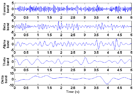
EEG signals differ based on electrode placement and brain activity. To interpret them, they are commonly divided into distinct frequency bands, each named after a Greek letter, and each associated with specific mental states.
- Delta (<3 Hz): Prominent in deep sleep.
- Theta (3–8 Hz): Associated with light sleep and drowsiness.
- Alpha (8–13 Hz): Seen when relaxed with eyes closed, representing synchronized brain activity.
- Beta (13–30 Hz): A Linked to alertness and active thinking, showing desynchronized patterns.
- Gamma (30–100 Hz): Linked to higher cognitive functions such as perception, memory processing, and consciousness.
Frequency-Domain Representation of EEG Signals
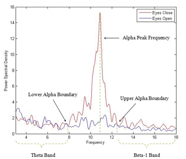
EEG signals can also be analyzed in the frequency domain using spectral analysis. The power spectral density (PSD) plot represents the distribution of signal power across different frequency bands. Figure 7 shows a typical PSD of EEG signals recorded from an awake resting subject under eyes-open and eyes-closed conditions. It can be observed that alpha band activity (8–13 Hz) is prominent during eyes-closed condition and gets suppressed when the eyes are opened. This demonstrates the dependence of EEG rhythms on physiological and mental states.
EEG Electrodes Placement and Acquisition
EEG can be recorded by three types of electrodes:
- EEG — Scalp — Outer Surface (Over Scalp)
- ECoG — Cortical — Exposed Surface
- Depth Records — Depth — In Deep Brain
- Wet electrodes (gel): Provide low impedance and high-quality recordings, standard in clinical use.
- Dry electrodes:Faster setup and portable, but with higher impedance and lower accuracy.
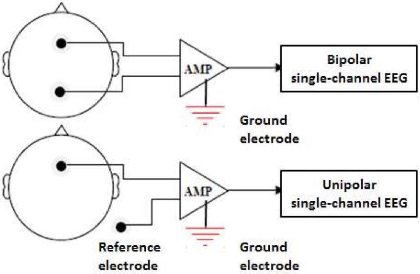
EEG can be recorded in two modes (Fig.8):
- Unipolar (monopolar)
- Bipolar
10–20 EEG Electrode Configuration
Placement of electrodes on the scalp is commonly dictated by the requirements of the measurement to be made. In clinical practice, a standard pattern, called the 10–20 electrode placement system, is generally used. This system, devised by a committee of the International Federation of Societies for Electroencephalography, is so named because electrode spacing is based on intervals of 10 and 20 percent of the distance between specified points on the scalp. Position is based on distance between Nasion to Inion i.e. from front to back and distance between two ear lobes. The 10–20 EEG electrode configuration is illustrated in Fig:8.
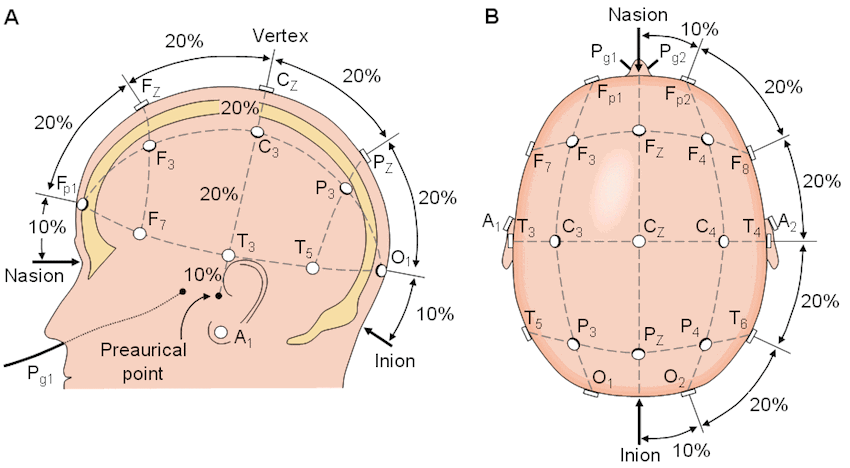
In all there are 21 electrodes: 19 active electrodes and 2 reference electrodes –
- Prefrontal: Fp1, Fp2
- Frontal: Fz, F3, F4, F7, F8
- Central: Cz, C3, C4
- Occipital: O1, O2
- Parietal: Pz, P3, P4
- Temporal: T3, T4, T5, T6
- Earlobes (reference): A1, A2
Applications of EEG Acquisition
EEG acquisition systems are used in numerous clinical and research applications:
- Clinical diagnosis: Identification of seizures, epilepsy, sleep disorders, and brain tumor.
- Neuroscience research: Investigation of cognitive processes, attention, and memory.
- Brain-computer interfaces (BCIs): Real-time decoding of brain signals for prosthetic control.
- Neurological monitoring: Continuous assessment of brain function during anesthesia or critical illness.