
Acquisition of EMG signals using biomedical systems
Procedure for EMG Acquisition
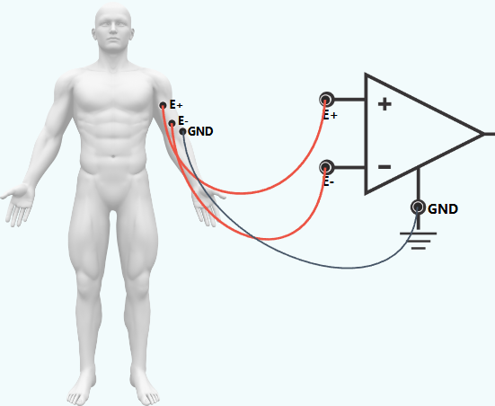
1. from Biceps (Elbow Flexion):
Step 1: Establish connections
- Connect the E+ electrode on Biceps to the positive input (E+) of the op-amp circuit.
- Connect the E- electrode on Biceps to the negative terminal (E-) of first op-amp circuit.
- Connect the GRD electrode nearby bony landmark to the ground (GRD) of the op-amp circuit.
Step 2:
Refer to the accompanying figure for correct connection points.
Step 3:
Click the Check button to verify correct connections.
Step 4:
Click the show signal button to display the simulated EMG waveform.
Step 5: Click the Normal button to observe the baseline EMG signal when the muscle is relaxed. Then click the Contraction button to simulate muscle Contraction and observe the increase in EMG amplitude.
Step 6:
Click the Signal button to pause the displayed EMG waveform.
Step 7:
Click the Print button to save the record observations.
Step 8: Select another muscle from the left panel to record EMG Signals for its corresponding muscle.
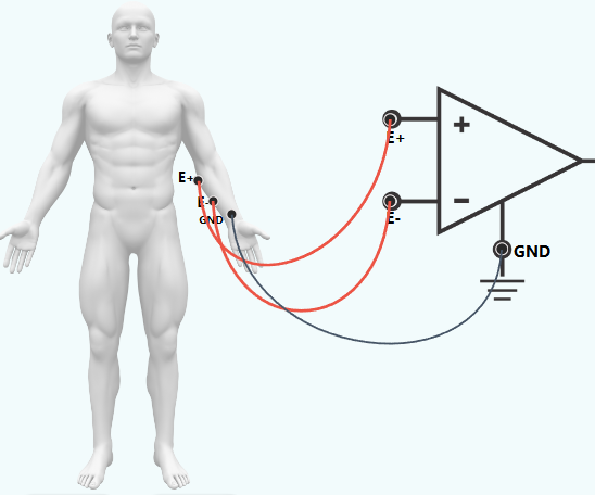
2.EMG Acquisition from Forearm Flexors (Grip):
Step 1: Establish connections
- Connect the E+ electrode on Forearm Flexors to the positive input (E+) of the op-amp circuit.
- Connect the E- electrode on Forearm Flexors to the negative terminal (E-) of first op-amp circuit.
- Connect the GRD electrode nearby bony landmark to the ground (GRD) of the op-amp circuit.
Step 2:
Refer to the accompanying figure for correct connection points.
Step 3:
Click the Check button to verify correct connections.
Step 4:
Click the show signal button to display the simulated EMG waveform.
Step 5: Click the Normal button to observe the baseline EMG signal when the muscle is relaxed. Then click the Contraction button to simulate muscle Contraction and observe the increase in EMG amplitude.
Step 6:
Click the Signal button to pause the displayed EMG waveform.
Step 7:
Click the Print button to save the record observations.
Step 8: Select another muscle from the left panel to record EMG Signals for its corresponding muscle.
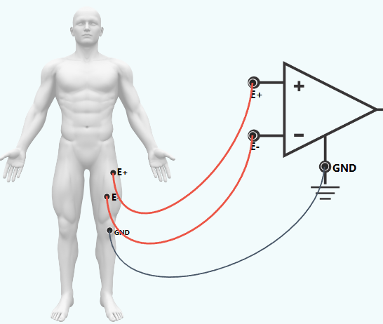
3.EMG Acquisition from Quadriceps (Knee Extension):
Step 1: Establish connections
- Connect the E+ electrode on Quadriceps to the positive input (E+) of the op-amp circuit.
- Connect the E- electrode on Quadriceps to the negative terminal (E-) of first op-amp circuit.
- Connect the GRD electrode nearby bony landmark to the ground (GRD) of the op-amp circuit.
Step 2:
Refer to the accompanying figure for correct connection points.
Step 3:
Click the Check button to verify correct connections.
Step 4:
Click the show signal button to display the simulated EMG waveform.
Step 5: Click the Normal button to observe the baseline EMG signal when the muscle is relaxed. Then click the Contraction button to simulate muscle Contraction and observe the increase in EMG amplitude.
Step 6:
Click the Signal button to pause the displayed EMG waveform.
Step 7:
Click the Print button to save the record observations.
Step 8: Select another muscle from the left panel to record EMG Signals for its corresponding muscle.
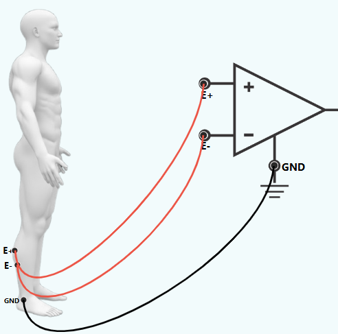
4.EMG Acquisition from Gastrocnemius (Calf Raise):
Step 1: Establish connections
- Connect the E+ electrode on Gastrocnemius to the positive input (E+) of the op-amp circuit.
- Connect the E- electrode on Gastrocnemius to the negative terminal (E-) of first op-amp circuit.
- Connect the GRD electrode nearby bony landmark to the ground (GRD) of the op-amp circuit.
Step 2:
Refer to the accompanying figure for correct connection points.
Step 3:
Click the Check button to verify correct connections.
Step 4:
Click the show signal button to display the simulated EMG waveform.
Step 5: Click the Normal button to observe the baseline EMG signal when the muscle is relaxed. Then click the Contraction button to simulate muscle Contraction and observe the increase in EMG amplitude.
Step 6:
Click the Signal button to pause the displayed EMG waveform.
Step 7:
Click the Print button to save the record observations.
Step 8: Select another muscle from the left panel to record EMG Signals for its corresponding muscle.